
However, not all congenital anomalies can be detected by sonography.
Some congenital anomalies may also develop later during pregnancy.
The ability to detect congenital anomalies depends on the amount of amniotic fluid, gestational age, and fetal position.
By sonography, approximately 45% to 85% of anomalies can be detected, depending on the gestational age.
Sonography performed before 18 weeks or after 30 weeks of gestation is not appropriate for detailed anomaly evaluation.
The following anomalies cannot be detected with certainty by this sonography:
- Bicuspid aortic valve
- Small ventricular septal defect (VSD) in the heart
- Small isolated septal defects
- Isolated cleft palate
- Exact number of fingers and toes
- Ear anomalies
- Functional abnormalities such as hearing or visual impairment
- Intellectual disability
- Oesophageal atresia
- Anal atresia
- Certain congenital anomalies
- All metabolic diseases
- Genetic disorders
- Syndromes
- Mutation-related abnormalities
- Ambiguous genitalia

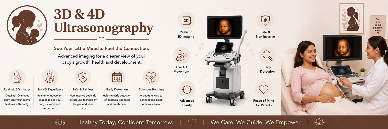
3D and 4D ultrasound of the fetus are advanced medical imaging technologies used during pregnancy to view the baby’s development and well being more closely. Compared with standard (2D) ultrasound, these techniques can be very helpful for both parents and doctors. Below is a complete scientific and medical explanation:
1. Difference between 2D, 3D and 4D ultrasound
These three methods used to examine the fetus are quite different:
- 2D ultrasound (conventional): The traditional method that produces black‑and‑white flat images of the baby’s internal organs and bones. Doctors mainly use this for internal anatomical assessment.
- 3D ultrasound (three‑dimensional): This technology combines length, width and depth to produce a still, color-like three‑dimensional image of the baby’s face and body. The baby appears more like a real photograph.
- 4D ultrasound (four‑dimensional): This is essentially 3D plus the fourth dimension — time. It provides live video, so parents can watch the baby move in real time (yawning, sucking thumb, smiling, etc.).
2. When to do 3D 4D
ultrasound
These scans can be performed at any stage of pregnancy, but the ideal window to clearly see facial features and limbs is between 20 and 32 weeks (5–8 months):
- 20–26 weeks: At this stage there is less subcutaneous fat under the baby’s skin, so bone structure appears very clear.
- 27–32 weeks: The baby’s facial features (eyes, nose, lips) are well developed and the face appears very clear. After about 32 weeks, the baby is larger and there is less space in the uterus, which can make viewing the face more difficult.
3. Medical benefits of this ultrasound
Many people use 3D‑4D scans primarily to see the baby’s face, but there are important clinical advantages:
- Early detection of congenital anomalies: External defects such as cleft lip/palate or limb malformations can be detected earlier and more accurately with 3D imaging.
- Assessment of face and skull: Provides detailed information about cranial bone and facial structure development.
- Spine evaluation: Very accurate for detecting vertebral abnormalities (e.g., spina bifida).
- Placenta and cord position: More clearly shows placenta location and whether the umbilical cord is wrapped around the baby’s neck.
4. Is 3D 4D ultrasound safe?
- Yes. When performed by a qualified radiologist or gynecologist, these scans are considered safe for both mother and baby.
- Ultrasound uses high‑frequency sound waves, not ionizing radiation like X‑rays. However, medical guidelines advise avoiding repeated or prolonged scans done only for souvenirs or videos; scans should be performed on medical indication and doctor’s recommendation.
5. Limitations
Even with 3D‑4D ultrasound, images or videos may not be clear due to:
- Fetal position: If the baby’s face is turned toward the mother’s spine (facing backward), the face may not be visible.
- Amniotic fluid volume: Low fluid around the fetus reduces image quality.
- Maternal body habitus: Excess fat in the abdominal wall (obesity) can impede sound wave penetration and reduce image clarity.
Conclusion: 3D‑4D ultrasound is a wonderful way for parents to form an emotional bond with their unborn baby and is also a precise clinical tool for doctors to ensure fetal health.
